
Thanks to the ease of communication today, dozens of patients write to us daily through email, WhatsApp, Instagram, Facebook, and similar channels, describing their complaints and sending us a few test results, expecting advice and answers.
Can a doctor really make a diagnosis this easily? Unfortunately, no. The eye is our most precious and complex organ.
There are many different parts of our eyes, and these parts can be affected by hundreds, even thousands, of different diseases. Therefore, it is impossible to diagnose and plan treatment for eye diseases without a detailed examination and advanced tests.
In an eye examination, we, as ophthalmologists, start by measuring visual acuity and proceed to examine many areas, such as the eyelids, conjunctiva, cornea, anterior chamber, iris, lens, vitreous, retina, optic nerve, and visual pathways leading to the brain, while also measuring the function of these areas.
In short, although it may seem like a simple eyeglass examination, we are looking for clues to hundreds or even thousands of diseases during the examination and making diagnoses accordingly. Additionally, an eye examination is one of the most equipment-dependent exams in medicine. Dozens of different high-tech devices are used to visualize and measure the functions of the aforementioned areas. In this article, I will try to explain the steps of an eye examination to you.
An eye examination is the process of anatomically evaluating the eye to check whether all its parts are healthy and measuring visual functions with tests to determine whether they are normal.
For example:
Most of our patients understand this when they think of an eye exam. It is the stage where we ask patients to read letters from a distance. This helps us understand how well the patient can see at a certain distance. Before this, we measure the patient’s refractive error with an auto-refractometer (computerized eye exam machine).
If the patient cannot read all the letters from a distance without glasses, we move to the second stage, which is the eyeglass prescription exam, to see if they can read with glasses.
The high-tech devices we use measure the eyeglass prescription for most patients. We learn the values of refractive errors such as myopia, hyperopia, and astigmatism. However, these objective measurements don’t always guarantee good vision.
First of all, if there is an underlying disease such as glaucoma, cataracts, or retinal disease, glasses won’t improve the patient’s vision, as glasses only help focus the image onto the retina. In other words, glasses correct refractive errors, which are adjustments in the eye’s focusing mechanism when there is no disease.
For instance, in myopia, if the image is focused in front of the retina, the lenses we prescribe shift the focus onto the retina. However, if there is a problem with the retina, the patient still won’t see clearly with glasses because even if the focus is correct, the eye's tissues must be healthy for vision to occur.
During the examination, the patient needs to be very attentive because the doctor doesn’t actually prescribe glasses but allows the patient to try different lenses and choose the best one. This step determines which lens provides the best vision for the patient.
If the patient is over 45 years old, they will likely have difficulty with near vision as well. At this stage, an additional prescription is added to their distance prescription to determine the correct reading glasses. We also provide reading cards to the patient, and we determine the best near vision prescription based on the distance at which they want to read.
Many patients are ready to get up from the chair at this point, but the eye examination has not yet begun. What’s completed so far is just the vision and eyeglass examination.
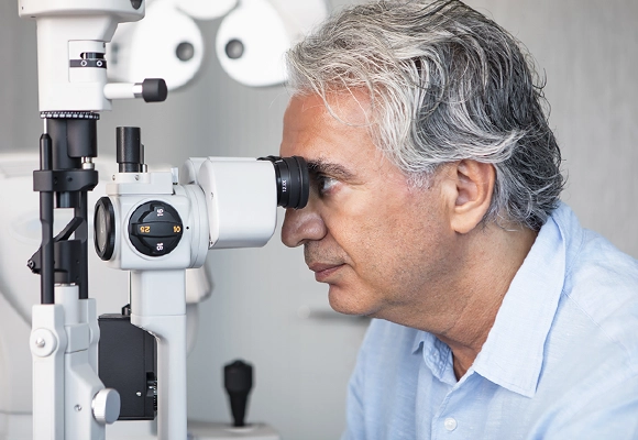
We call the examination of the eyelids, eyelashes, meibomian glands, conjunctiva, cornea, anterior chamber, iris, and lens the "anterior segment examination." Using a special microscope, we examine the patient's eye to investigate diseases in these areas.
When an experienced ophthalmologist looks at the eye with this device called a biomicroscope, they can instantly diagnose hundreds or even thousands of diseases. Many diseases, from dry eyes to uveitis, cataracts to glaucoma, can be understood by looking at the biomicroscope. This device also performs tear tests, eye pressure measurements and retinal examinations with the help of special lenses.
Intraocular pressure (IOP) can be measured using various devices. The most commonly used device is the air puff tonometer, which is known for blowing air into the eye. While this method is practical, it is not particularly reliable for glaucoma patients.
The most accurate method is performed by the doctor using a blue light on a slit lamp (biomicroscope). For this reason, this method should be preferred, especially in glaucoma patients during an eye examination.
The retina, also known as the neural layer, lines the inside of the eye and is responsible for capturing visual information. The image focused on the retina, particularly on the macula (the central area),is perceived by photoreceptor cells, which send electrical impulses to the optic nerve, leading to visual perception in the brain.
A retinal examination has several stages. First, the central area can be observed using special lenses. To examine the peripheral areas of the retina, the pupil may need to be dilated with drops. For macular examination, the OCT (Optical Coherence Tomography) device is crucial, as it reveals defects in the lower layers that cannot be seen with the naked eye, making it one of the most important examination tools.
Prof. Dr. Ahmet Akman personally performs the OCT test using the latest technology on every patient he examines, for both macular and optic nerve evaluation. Although OCT is often referred to as "eye tomography," it actually does not involve radiation. Tomography refers to three-dimensional imaging, and the tomography devices that use radiation are found in the radiology department. Eye tomography, on the other hand, uses light to map the back of the eye and contains no radiation.
In retinal examinations, in addition to diagnosing macular diseases, hundreds of conditions can be identified, including retinal vein occlusions, retinal tears, and diabetic retinopathy.
Today, OCT is indispensable for every eye examination. It should be as standard as an EKG during a cardiac exam.
Another part of the posterior segment examination is the optic nerve examination. By assessing the condition of the optic nerve and imaging it with the OCT device, it is possible to diagnose optic nerve inflammations, glaucoma damage, and even brain tumors.
In our clinic, we examine every patient's optic nerve using the OCT device to investigate any potential diseases. Optic nerve tomography is also personally performed by Prof. Dr. Ahmet Akman.
Following these standard examinations, 99% of the patient's issues are typically understood. The next step is to move on to specialized examination methods and tests tailored to the specific problem.
If the patient cannot see well even with glasses during the vision test and the doctor detects the presence of cataracts in the anterior segment examination, various measurements of the eye are taken using special devices. The standard examination steps are completed, and the pupil is dilated.
If the cataract is at a stage requiring surgery, the lens power to be placed in the eye is measured with a biometry device. The type of lens suitable for the patient is discussed, and the surgery planning begins.
In addition to the standard eye examination steps, the optic nerve and macula are examined using OCT eye tomography to check for any damage. If OCT reveals damage, a visual field test is conducted to assess the patient's functional loss.
To determine the type of glaucoma, an angle examination called gonioscopy is performed, which evaluates the drainage channels of the eye fluid, distinguishing between open-angle glaucoma and closed-angle glaucoma.
If the patient's symptoms point to dry eye, the tear film evaluation begins with the anterior segment examination. The presence of blepharitis and issues with the meibomian glands are investigated, and the lipid layer of the tear film is checked. Next, different tests are used to measure the aqueous layer of the tear film, and a treatment plan is made.
In addition to a standard retina examination, an OCT scan of the macula is performed. If bleeding is observed during the examination, an FFA test, also known as a fluorescein angiography, is applied. Based on these results, a treatment plan is created. In addition to the diseases briefly mentioned here, many different tests are used to diagnose and plan the treatment of other eye diseases.
Tests are conducted based on the nature of the disease and the patient's complaints. The main tests include:
Eye drops are required to dilate the pupils during a retina examination and especially during vision tests for children. Additionally, numbing drops are used for measuring eye pressure and in cases where the front part of the eye needs to be touched.
Patients with complaints should have an eye examination immediately. For individuals with a family history of serious eye diseases, frequent check-ups are necessary as per the doctor's recommendation. However, for individuals without any symptoms, regular eye exams are essential for the early diagnosis of many conditions.
The recommended schedule and frequency for eye examinations are as follows:
Didn't find what you need? Contact us today.
Contact Us Perfect Vision Turkey
Très bonne expérience avec le Docteur Safiye Yilmaz. Je leur ai expliqué que je devais retourner en France le Vendredi donc j’ai réussi à avoir un rendez-vous Mardi. La c{...}
26.03.2024Ich bin mit der gesamten Abwicklung vom ersten Termin bis hin zu der OP super zufrieden. Wer sich für eine Trifokallinse entschieden hat ist bei Frau Prof. Dr. Safiye Yil{...}
22.03.2024Uso gli occhiali da anni, finalmente me ne sono sbarazzato, sono molto soddisfatto, grazie mille / I've been using glasses for years, I finally got rid of it, I'm very sa{...}
21.03.2024